21 Luxury Human Liver Cell Diagram
Human Liver Cell Diagram esophagus American English or oesophagus British English s f s commonly known as the food pipe or gullet gut is an organ in vertebrates through which food passes aided by peristaltic contractions from the pharynx to the stomach The esophagus is a fibromuscular tube about 25 centimetres long in adults which travels behind the trachea and heart passes Human Liver Cell Diagram innerbody image card01 htmlThe heart is a muscular organ about the size of a closed fist that functions as the body s circulatory pump It takes in deoxygenated blood through the veins and delivers it to the lungs for oxygenation before pumping it into the various arteries which provide oxygen and nutrients to body tissues by transporting the blood throughout the body
cloning is the creation of a genetically identical copy or clone of a human The term is generally used to refer to artificial human cloning which is the reproduction of human cells and tissue It does not refer to the natural conception and delivery of identical twins The possibility of human cloning has raised controversies These ethical concerns have prompted several nations to pass Human Liver Cell Diagram bbc uk science humanbody body factfiles liver liver shtmlFeb 12 2004 Largest internal organ Your liver is your largest internal organ A big blood vessel called the portal vein carries nutrient rich blood from your small intestine directly to your liver myscience8 human biology digestive system lab 2007 pdfGiant Food Processor pieces in the mouth Carbohydrates The Digestive System is a Mechanical Digestion Food is chopped and ground into small
womens health advice human body htmlLIVER Click to enlarge Location Under the diaphragm slightly to the right side of the body Function To rid toxins from the blood to control blood sugar and produce bile for digestion purposes Things That Can Go Wrong Cirrhosis Liver damage usually caused by either longterm alcohol abuse or hepatitis C Human Liver Cell Diagram myscience8 human biology digestive system lab 2007 pdfGiant Food Processor pieces in the mouth Carbohydrates The Digestive System is a Mechanical Digestion Food is chopped and ground into small people eku edu ritchisong RITCHISO 301notes1 htmThere are two types of cells that make up all living things on earth prokaryotic and eukaryotic Prokaryotic cells like bacteria have no nucleus while eukaryotic cells like those of the human body do So a human cell is enclosed by a cell or plasma membrane Enclosed by that membrane is the cytoplasm with associated organelles plus a nucleus
Human Liver Cell Diagram Gallery

600px Liver_histology_005, image source: embryology.med.unsw.edu.au
circulatory system diagram labeled human circulatory system simple, image source: pixshark.com

image4, image source: www.pancreapedia.org

Cirrhosis of the Liver, image source: www.ezhealthmd.com

histology of gastrointestinal tract 44 638, image source: www.slideshare.net

KidneyAnatomy, image source: sites.google.com
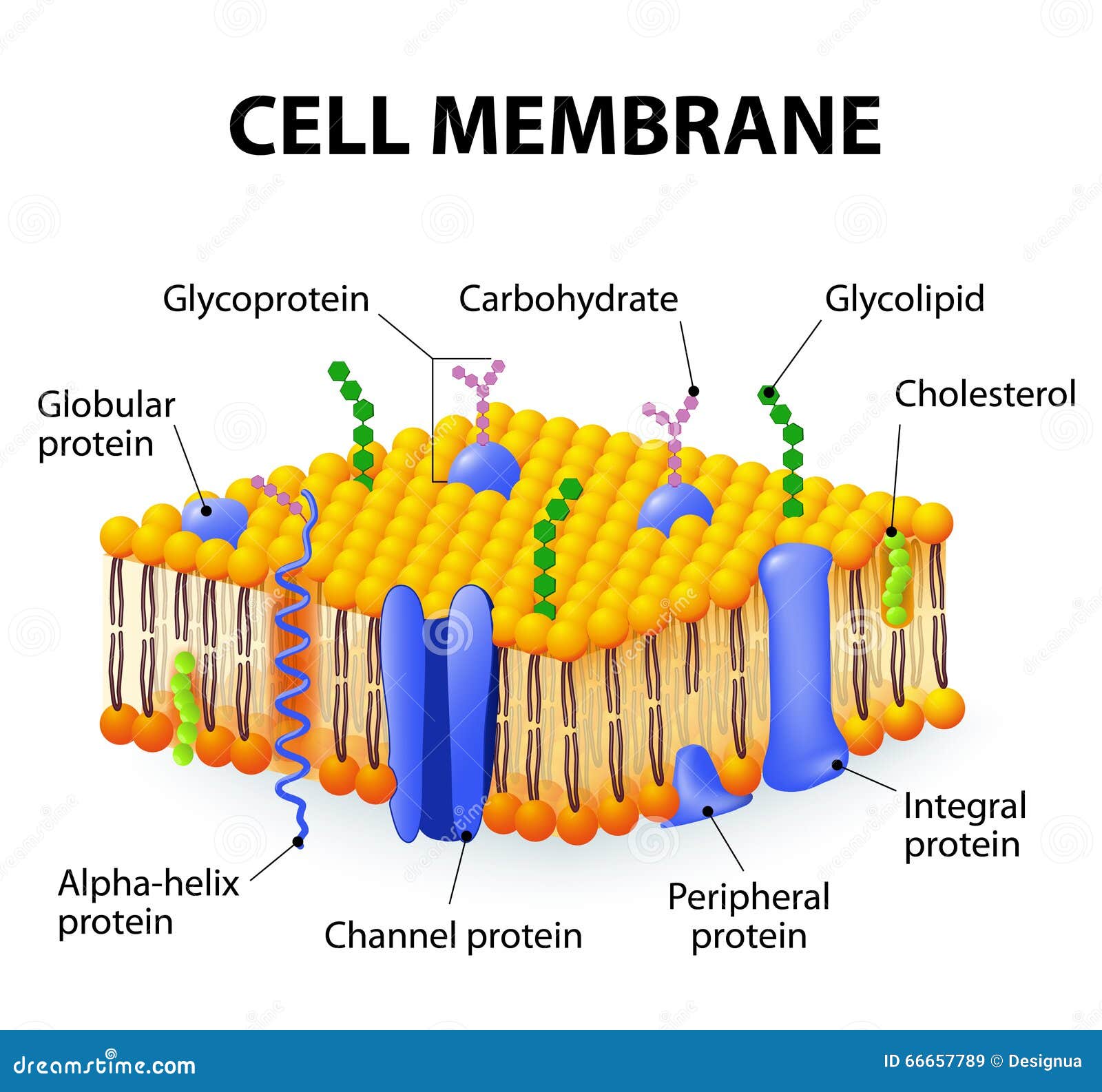
cell membrane detailed diagram models structure 66657789, image source: www.dreamstime.com

TOMCJ 8 28_F2, image source: benthamopen.com


2105_Capillary_Bed, image source: www.lookfordiagnosis.com
e coli, image source: sgugenetics.pbworks.com

hqdefault, image source: www.youtube.com
path blood takes, image source: www.edinformatics.com

bigstock stem cells 74626423, image source: www.askdrray.com

stages of liver damage due to alcoholism first alcohol compromises G156NE, image source: www.alamy.com


Pancreas Pictures 3, image source: www.fbs-wp.leeds.ac.uk

western blot procedure schematic, image source: www.sigmaaldrich.com

afp20050101p105 f1, image source: www.aafp.org
aerobic and anaerobic proce_med, image source: ib.bioninja.com.au


malaria_lifecycle, image source: blogusscientificus.blogspot.com
Meniscus Tears Color 1, image source: elsalvadorla.org


Comments
Post a Comment